Diagnostics & Imaging
OCT, Fundus and Visual Field Integration: EMR Checklist
Use this imaging EMR checklist to evaluate patient matching, laterality, modality support, file validation, review, comparison, security, and recovery.
In this article
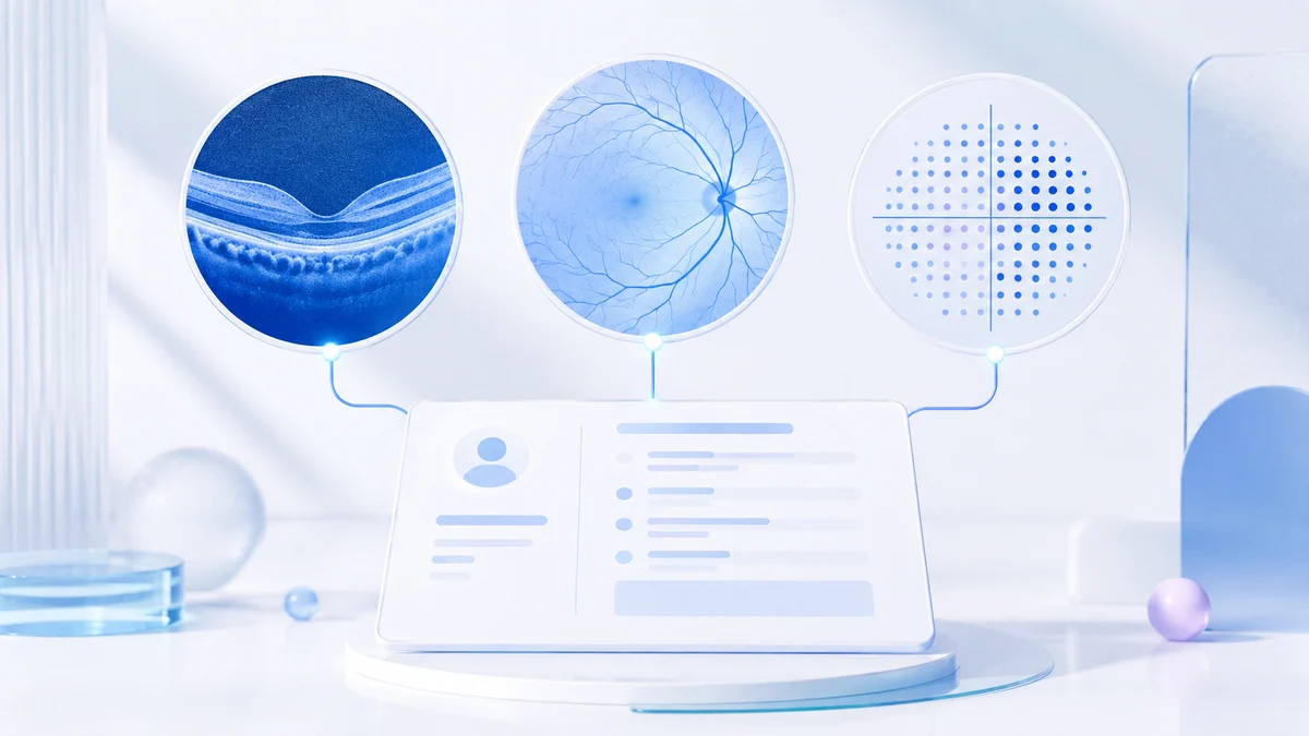
Ophthalmic imaging integration should connect the correct patient, encounter, eye, modality, report, image, technician action, and clinician review. A vendor saying “we support DICOM” is only the beginning of the evaluation.
Key takeaways:
- Test each device and data path separately.
- Preserve patient identity, date, modality, and laterality.
- Distinguish acquisition, completion, and clinician review.
- Include failure recovery, access control, and longitudinal comparison.
Identity and encounter matching
Confirm how the modality receives patient and appointment identity. Test duplicate names, changed demographics, emergency patients, corrected identifiers, and uploads made after the original visit. Avoid workflows that depend on staff retyping identifiers at every device.
Laterality and modality
Verify that OD, OS, and OU are stored explicitly rather than inferred from filenames. Confirm support for the exact OCT, fundus, visual field, biometry, topography, pachymetry, and other systems used by the hospital.
File and report controls
Ask which formats are supported, how size and type are validated, where files are stored, how thumbnails and reports are generated, and how failed or partial uploads are handled. Confirm that a report cannot be attached to the wrong tenant or branch.
Technician completion and clinician review
Acquiring an image is not the same as completing a diagnostic workflow, and completion is not the same as doctor review. The system should preserve who performed each action and route review appropriately.
Longitudinal comparison
Open several dated studies and assess side-by-side comparison, laterality, report text, image quality, measurement trends, and visit context. AI summaries should cite the saved source encounters and surface missing or conflicting evidence.
Integration acceptance checklist
- Exact device and software version tested
- Patient and encounter identity verified
- OD/OS/OU confirmed
- Original image and report retained
- Upload interruption recovered safely
- Unauthorized branch access rejected
- Technician and reviewer actions attributable
- Historical studies accessible without entering live queues
Distinguish file upload from device integration
| Connection | What it may provide | What still needs testing |
|---|---|---|
| Manual image or PDF upload | A report attached to the record | Identity checks, laterality and version history |
| Device export or watched folder | Transfer with less manual copying | Duplicate handling, failure alerts and patient matching |
| DICOM connection | Agreed imaging objects and exchange services | Supported services, formats and device conformance |
| Structured measurement interface | Values available for search or trends | Units, eye, dates and meaning of each field |
An attached PDF may be useful without making its measurements available for a trend. Ask the vendor to show both the original report and any extracted values, so the team can tell which information was transferred and which was interpreted.
The DICOM conformance guidance calls for validation between the specific systems being connected. Obtain the device manufacturer's conformance statement and test an interrupted transfer, a repeated upload and an identifier correction before accepting the connection.
For existing archives, use the separate EMR data migration checklist. Integration of future studies and migration of old studies need their own acceptance checks.
Frequently asked questions
Is DICOM support enough to guarantee integration?
No. Verify the exact modality, conformance, worklist, network, patient-matching, report, image, and support workflow.
Should a doctor’s advice create the imaging workflow automatically?
Not in Iris. Consultation advice remains in the clinical plan until the hospital deliberately initiates its operational workflow.